Finding explained
They saw a bright spot in the heart. Here is what it means.
The report says "echogenic intracardiac focus." Three long words for something visually dramatic but usually ordinary: a small spot that reflects ultrasound the way bone does. On this page: what it is, the actual risk numbers, and what guidelines say to do about it.
The short answer
An echogenic intracardiac focus (EIF) is a bright dot inside a heart chamber, seen in 3-5% of normal pregnancies - up to 30% in some Asian populations. It is not a hole or a structural defect. When it is the only finding and your screening test for chromosome conditions was negative, guideline-issuing societies say no further testing is needed. About 90% disappear by the third trimester.
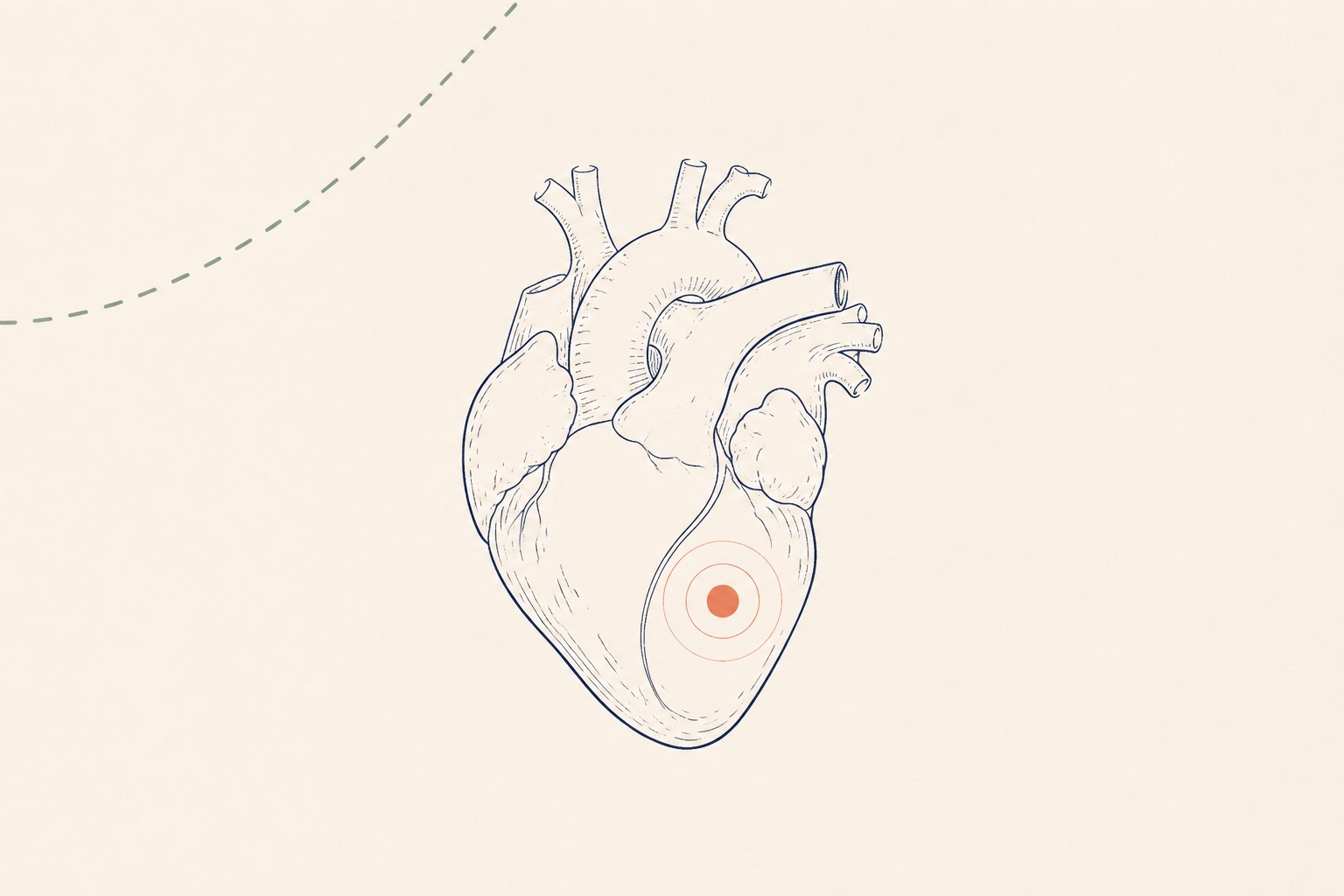
What is an EIF, physically?
Ultrasound sees density. When a small area inside the heart - usually a papillary muscle, the tiny muscle cord that anchors a heart valve - picks up calcium mineralization, it bounces sound back as brightly as bone. The machine paints it white. That is the entire phenomenon: a mineral fleck on an ultrasound image.
It is not a hole in the heart. It is not a valve leaking. It does not obstruct anything. Roughly 88% sit in the left ventricle; some are right-sided or bilateral.
Evidence
Definitions, left-ventricle predominance (88%), and resolution rates follow the clinical review of sonographic soft markers: J Genet Med 2018; ethnic prevalence data from the SMFM Consult Series #57 (AJOG 2021).
"Does it mean Down syndrome?" - the numbers, without flinching
This is the question that keeps people up after this finding, so it deserves actual numbers rather than "don't worry."
The largest combined analysis to date pooled over 104,000 pregnancies. An isolated EIF was present in 4.29% of chromosomally normal fetuses and 11.49% of fetuses with trisomy 21. That asymmetry is why EIF was ever called a "marker" at all - it shifts probability slightly upward.
But shift how much? A positive likelihood ratio of about 2.7 means: whatever your baseline odds were, multiply them by roughly two-and-a-half. If your baseline was 1 in 1,000 (typical after negative screening), you are now at roughly 1 in 400. Still far below the thresholds where anything is recommended.
Evidence
104,001 pregnancies; isolated ECF in 4,416/102,847 euploid fetuses (4.29%) vs 64/557 trisomy 21 fetuses (11.49%), LR+ 2.68: Ultrasound Int Open 2019.
What the guidelines actually recommend
The Society for Maternal-Fetal Medicine reviewed this question across every large study and issued a graded recommendation in 2021. Two situations cover almost everyone:
- You already had NIPT or serum screening, and it was negative: no further evaluation. The guideline's exact framing is that an isolated EIF in this situation is "a normal variant of no clinical importance" - no fetal echocardiogram, no follow-up scans, no postnatal testing (GRADE 1B).
- You have not had any chromosome screening this pregnancy: the recommendation is a counseling conversation about your baseline risk and the option of NIPT or a quad screen. Not automatic invasive testing - the numbers above are why.
If other findings appeared alongside the EIF - a thickened nuchal fold, echogenic bowel, structural differences elsewhere - then it is the combination that matters, and your clinicians will treat the total picture, not the spot.
Evidence
SMFM Consult Series #57: negative screening + isolated EIF → "no further evaluation... normal variant of no clinical importance" (GRADE 1B): AJOG 2021.
What happens next, concretely
- Check what screening you already had. If NIPT or a quad screen came back low-risk this pregnancy, the file on an isolated EIF is essentially closed.
- If you have not screened yet, an NIPT conversation is the one decision this finding may prompt. It is a maternal blood draw, carries no miscarriage risk, and re-sorts the odds with high accuracy for trisomy 21.
- Ask whether anything else was noted - on the heart views or anywhere else. "Isolated" is the load-bearing word in every statistic above.
- Let the spot go. Roughly 90% are gone from view by the third trimester. Even the ones that stay do not, by themselves, change anything.
When the fear does not close the file
Some parents read all of this and still cannot sleep. That is not a reasoning failure - it is how anxiety works around prenatal findings. Two things tend to help more than re-reading statistics:
First, put the actual words of your report in front of your clinician and ask them to say, out loud, what they would do if this were their own scan. Second, if the waiting itself has become the problem, the waiting-days guide covers how to get through the gap between a finding and the next appointment without catastrophizing at 2 a.m.
Bring these to your next appointment
- Is the EIF isolated, or were other findings noted on any view?
- Given my screening result, does this change any recommendation for me?
- Do I need a fetal echocardiogram for this finding alone?
- Where in the heart is it, and does that matter in my case?
- Will anyone look again later in pregnancy either way?