Evidence update · checked 2026-10-28
Suspected coarctation on fetal echo: why over half of flagged babies do not need arch surgery
Few phrases on a fetal echocardiography report unsettle families quite like "cannot exclude coarctation of the aorta." You are told the baby may need heart surgery in the first week of life, and in the same breath you are told the heart might turn out to be completely unobstructed once the baby is born. That contradiction is not hedging by an uncertain doctor. It is a structural feature of how fetal circulation works before birth, and recent multi-year cohorts from Genoa, Zhejiang, and North American fetal heart programs finally put clear numbers on both sides of the coin.
The short answer
Across published cohorts, 45% to 70% of fetuses flagged for suspected coarctation do not require aortic arch surgery after birth. Before delivery, the open ductus arteriosus masks how the aortic isthmus will behave. Newer third-trimester Z-scores, arch angles, and strain metrics sharpen prenatal prediction, while a 48 to 72 hour postnatal hospital watch safely catches true cases.
Why the fetal arch hides its final shape until birth
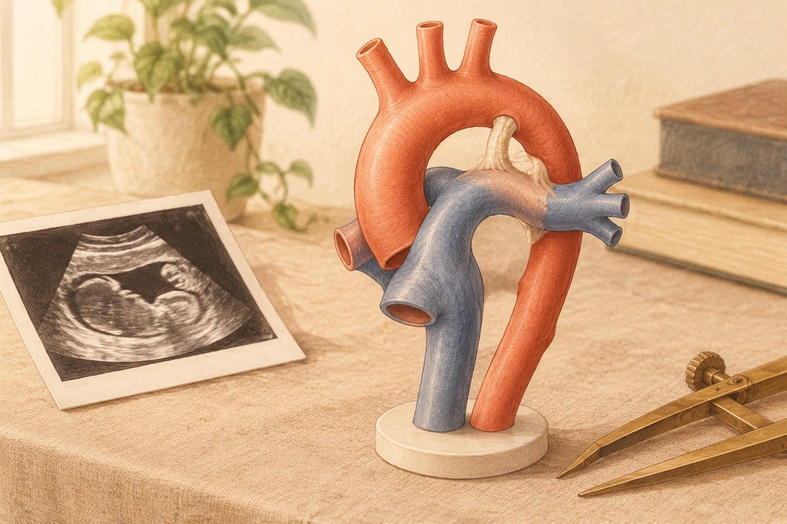
Coarctation of the aorta (CoA) is a narrowing of the upper descending aorta, almost always located at the aortic isthmus just where the ductus arteriosus joins the arch. In a newborn or child, a tight narrowing creates an obvious pressure step-up and high-velocity jet on Doppler ultrasound. In a fetus, that pressure gradient is absent.
During pregnancy, the lungs are filled with fluid and the wide-open ductus arteriosus routes most of the right ventricle's blood directly into the descending aorta. Only about 10% to 15% of combined cardiac output crosses the narrow aortic isthmus. Because the isthmus carries relatively little blood in utero, it naturally looks narrower than the pulmonary trunk, and when the right ventricle looks larger than the left (ventricular disproportion), sonographers flag suspected coarctation so a newborn is never sent home with an unrecognized ductal-dependent lesion.
What the 2022 to 2026 cohorts show
Three complementary studies show how the numbers shift depending on why the scan raised a flag and which measurements the fetal cardiology team uses:
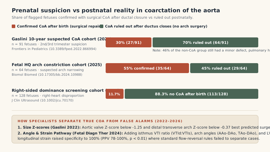
- The 10-year Gaslini Children's Hospital series (91 fetuses): Reviewing a decade of pregnancies referred for suspected CoA in the second or third trimester, investigators found that 27 of 91 babies (30%) were confirmed with coarctation after birth and underwent surgical repair, while 64 of 91 (70%) did not develop coarctation.
- The Fetal HQ arch-constriction cohort (64 fetuses): In a 2025 analysis of 64 fetuses with direct ultrasound suspicion of aortic arch constriction at Sir Run Run Shaw Hospital, 35 cases (55%) were confirmed postnatally and 29 cases (45%) were ruled out after birth.
- The 2026 perinatal right-dominance cohort (128 fetuses): When screening started from right-sided heart dominance across 128 fetuses meeting inclusion criteria, only 15 babies (11.7%) developed true postnatal coarctation; in those 15, the pulmonary-to-aortic valve (PV/AV) diameter ratio and the aortic isthmus Z-score were the strongest early markers.
Which echo measurements separate true CoA from false alarms?
For years, clinicians relied on qualitative signs such as right-to-left ventricular size discrepancy, a persistent left superior vena cava (PLSVC), or flow reversal across the foramen ovale. Recent pathway studies show why those older rules generated so many false alarms, and what works better on a third-trimester fetal echocardiogram:
| Echo metric or clinical sign | Cutoff or pathway finding | What published data show |
|---|---|---|
| Aortic valve (AV) & distal transverse arch (TAA) Z-scores | AV Z-score below -1.25; distal TAA Z-score below -0.37 |
Best third-trimester anatomical predictors in the 91-fetus Gaslini cohort; direct arch hypoplasia outperforms ventricular ratio alone. |
| Pulmonary-to-aortic valve ratio (PV/AV) & isthmus Z-score | Elevated PV/AV ratio with low aortic isthmus Z-score | Most reliable early-gestation parameter in the 2026 cohort of 128 right-dominant fetuses (80% of confirmed cases showed early signs). |
| Isthmus VTI ratio (VTId:VTIs), arch angles (AAo-DAo, TAo-DAo) & LV strain | Combined 4-metric pathway in moderate/high-risk fetuses | Separated surgical from non-surgical cases (p < 0.01 for all four) in the 2024 pathway study, achieving 100% specificity and 78% to 100% positive predictive value. |
| Redundant foramen ovale membrane or isolated PLSVC | Often noted on routine reports | Did not correlate significantly with confirmed postnatal CoA in the Gaslini multivariable analysis when arch Z-scores were normal. |
In the 2024 clinical pathway study published in Fetal Diagnosis and Therapy, standard high-risk criteria (such as flow reversal at the foramen ovale or aortic arch) failed to separate surgical from non-surgical babies within the moderate- and high-risk group. Adding four quantitative measures, the diastolic-to-systolic velocity-time integral ratio at the aortic isthmus (VTId:VTIs), the ascending-to-descending aorta angle (AAo-DAo), the transverse-to-descending aorta angle (TAo-DAo), and left ventricular longitudinal strain (LVS), separated the two groups with 100% specificity.
Why the 48 to 72 hour postnatal watch still matters
If half to two-thirds of flagged babies never need arch surgery, parents understandably ask why delivery planning at a cardiac-capable hospital is still recommended. The cohorts answer that question with two numbers that rarely make it into internet forum threads:
- The ductal closure window takes 48 to 72 hours: A newborn with severe coarctation often looks completely pink and comfortable in the delivery room because the ductus arteriosus is still open. Narrowing only declaration-tests the circulation as the ductus constricts on day two or three of life. In a planned delivery, doctors watch four-limb blood pressures, pre- and post-ductal pulse oximetry, and serial neonatal echocardiograms before symptoms ever appear, starting prostaglandin E1 only if the arch truly obstructs.
- The 46% secondary-finding rule: In the Gaslini cohort, among the 64 babies whose aortic arches turned out to need no surgery, 46% were still diagnosed after birth with another previously unseen minor cardiac defect (such as a small ventricular septal defect or bicuspid aortic valve), transient neonatal pulmonary hypertension, or a genetic condition. Delivering where a pediatric cardiologist can examine the baby in the first days of life closes the loop safely even when the arch is clear.
Four questions to bring to your next fetal echo
- What are the actual Z-scores for the aortic valve, distal transverse arch, and aortic isthmus? Is the concern based on a small arch, or mainly on right-left ventricular size discrepancy?
- Does your center classify our baby as mild, moderate, or high risk for postnatal coarctation? Do you measure arch angles or isthmus VTI ratios on the third-trimester scan?
- What is the exact postnatal observation protocol for our risk tier? Will the baby stay with us or go to the NICU for umbilical line monitoring and serial echoes while the ductus closes over 48 to 72 hours?
- Should we still plan a normal vaginal delivery near the cardiac team? (As covered on our delivery page, suspected CoA changes where you deliver and how the baby is watched in the first three days, not the mode of delivery.)
Sources (checked 2026-10-28)
Four-metric fetal CoA clinical pathway (VTId:VTIs, AAo-DAo and TAo-DAo angles, LV longitudinal strain): Fetal Diagnosis and Therapy 2024 (Fetal Diagn Ther 2024). Ten-year Gaslini Children's Hospital cohort (91 fetuses, AV Z-score -1.25 and distal TAA Z-score -0.37 cutoffs, 46% secondary findings in non-CoA group): Frontiers in Pediatrics 2022 (Front Pediatr 2022). Early right-heart dominance cohort (128 fetuses, PV/AV ratio and isthmus Z-score): Journal of Clinical Ultrasound 2026 (J Clin Ultrasound 2026). Fetal HQ arch constriction cohort (64 fetuses): Biomolecules and Biomedicine 2025 (Biomol Biomed 2025). Perinatal circulation and ductal-dependent delivery planning: AHA Scientific Statement, Circulation (Donofrio et al., Circulation).