Evidence update · checked 2026-11-22
Persistent fetal bradycardia and Long QT: why 120 bpm still triggers ECGs
The short answer
When a structurally normal fetus shows persistent sinus bradycardia without maternal anti-Ro/La antibodies or blocked premature atrial beats, inherited Long QT syndrome (LQTS) is the leading underlying diagnosis. Because fetal heart rate normally changes across pregnancy, doctors evaluate gestational Z-scores (-2 SD) rather than a single 110 bpm cutoff, and screen both parents with ECGs.
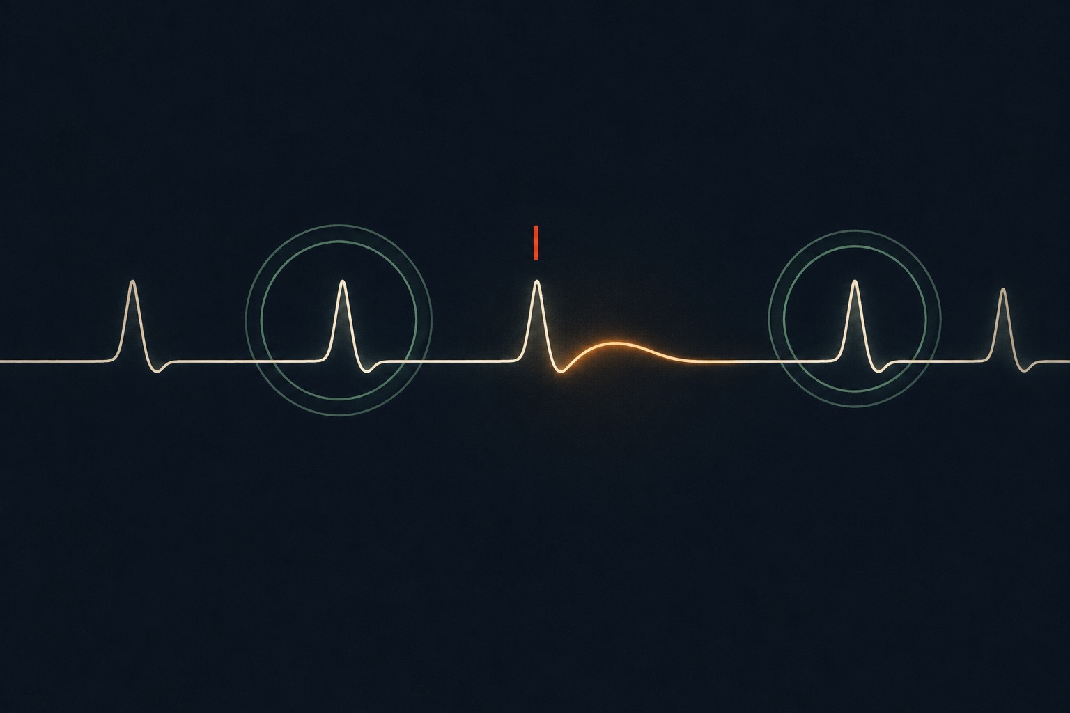
Parents preparing for a fetal echocardiogram rarely expect to hear about their own hearts. Yet when an obstetric ultrasound flags a consistently slow fetal heart rate, even numbers between 115 and 130 beats per minute in mid-pregnancy, pediatric electrophysiologists frequently hand both the mother and father orders for 12-lead electrocardiograms. Two recent clinical cohorts in Ultrasound in Obstetrics & Gynecology (2026) and Circulation: Arrhythmia and Electrophysiology (2025) explain why: persistent non-immune fetal bradycardia is frequently the first visible footprint of congenital Long QT syndrome (LQTS), and traditional textbook cutoffs miss many affected babies and carrier parents.
The counter-intuitive cutoff: why 120 bpm can still be fetal bradycardia
Most obstetric waiting-room handouts quote 110 beats per minute (bpm) as the hard lower limit of normal fetal heart rate. While 110 bpm works well as a labor-ward floor near term, it creates a dangerous blind spot in the second and early third trimesters, when a healthy fetus normally beats much faster (often 140 to 160 bpm around 20 weeks). Relying solely on a flat 110 bpm alarm misses babies whose heart rates sit consistently below the normal curve for their exact gestational week.
In a 2026 retrospective cohort from a tertiary fetal cardiology center published in Ultrasound in Obstetrics & Gynecology (Ultrasound Obstet Gynecol 2026, PMID 41482982), clinicians defined fetal bradycardia rigorously as a ventricular rate more than 2 standard deviations below the gestational-age-specific mean (FHR Z-score below -2.0). After excluding maternal anti-Ro/anti-La autoimmune heart block, blocked atrial ectopic beats, and major structural congenital heart disease, 22 fetuses met inclusion criteria between January 2018 and November 2023: 16 presented with isolated sinus bradycardia, 2 with functional 2:1 atrioventricular block (AVB), 2 with both sinus bradycardia and 2:1 AVB, and 2 with polymorphic ventricular tachycardia (VT).
The presenting heart-rate numbers carry a striking lesson for parents reading an ultrasound portal during the waiting days: the median presenting fetal heart rate in sinus rhythm was 120 bpm (ranging from 100 all the way up to 139 bpm), which corresponded to a median FHR Z-score of -3.04 (range -4.98 to -0.91). In other words, more than half of the referred fetuses in sinus rhythm would have passed a naive "above 110 bpm" visual check on a screening sheet, even though their heart rates sat three standard deviations below healthy gestational peers.
Among the 14 fetuses in the 2026 UOG study who underwent genetic testing (12 of them prenatally), 13 cases (92.9%) carried a confirmed pathogenic LQTS variant: 8 in KCNQ1 (LQT1), 2 in KCNH2 (LQT2), 1 in KCNE1, 1 in SCN5A, and 1 in CALM2. Remarkably, none of the 22 families had any known family history of Long QT syndrome prior to the fetal echo referral.
Separating benign PACs from autoimmune block and Long QT syndrome
Hearing the word "bradycardia" or "arrhythmia" on an anatomy scan report does not automatically mean Long QT syndrome. Fetal cardiologists follow a structured triage sequence on the first specialist scan to separate common, transient rhythm variations from inherited channelopathies or autoimmune inflammation. Understanding which findings belong to which tier helps parents read their fetal echo report calmly and accurately.
| Fetal rhythm pattern on echo | Primary biological mechanism | Key diagnostic test / cutoff | Typical clinical pathway & prognosis |
|---|---|---|---|
| Blocked atrial ectopic beats (PACs) | Immature atrial electrical triggers reset the AV node intermittently, mimicking a slow pulse | M-mode / Doppler shows irregular atrial beats followed by non-conducted pauses | Most common benign cause of brief slow fetal pulse; usually resolves spontaneously before or soon after birth without medication. |
| Autoimmune AV block (anti-Ro / anti-La) | Maternal SSA/SSB antibodies cross the placenta and inflame fetal AV conduction tissue | Maternal anti-Ro (SSA) & anti-La (SSB) blood panel + AV mechanical PR interval | Requires close serial surveillance (see Slow Heart Registry NCT04559425) and prompt anti-inflammatory protocol if progressing. |
| Persistent sinus bradycardia (no CHD, Ro-negative) | Delayed ventricular repolarization in sinus node ion channels (KCNQ1, KCNH2, SCN5A) | FHR Z-score < -2.0 SD for gestational week (median 120 bpm / Z -3.04 in UOG 2026) | High yield for LQTS (13/14 tested in UOG 2026); triggers parental 12-lead ECGs, prenatal genetic panel, and QT-safe drug precautions. |
| Functional 2:1 AV block or polymorphic VT | Severely prolonged ventricular refractory period prevents every second atrial beat from conducting | Atrial rate normal (~140 bpm), ventricular rate half (~70 bpm), or bursts of rapid VT | Highest-acuity LQTS presentation (e.g., KCNH2, SCN5A, or CALM2); managed at a Level 4 cardiac delivery center with immediate beta-blocker and pacing readiness. |
The 2026 UOG investigators also examined Doppler-derived mechanical surrogates of the fetal QT interval, specifically left ventricular isovolumetric relaxation time (LVIRT) and normalized LVIRT (N-LVIRT). While LVIRT was persistently prolonged in 6 of the 13 genotype-positive fetuses and N-LVIRT in 2 of 13, neither index caught every confirmed case. Consequently, a normal Doppler LVIRT measurement does not clear a fetus with persistent Z-score bradycardia from needing targeted genetic counseling and family ECG review.
Why both parents get ECGs (and why a normal QTc under 400 ms does not clear a carrier)
The second major clinical insight comes from a 2025 study published in Circulation: Arrhythmia and Electrophysiology (Circ Arrhythm Electrophysiol 2025, PMID 41328558), which evaluated 20 mothers and 20 fathers of fetuses referred for persistent non-immune bradycardia (16 with sinus bradycardia and 4 with functional 2:1 AV block).
Comprehensive parental evaluation and genomic testing (completed in 12 mothers and 11 fathers) uncovered pathogenic LQTS variants in 11 fetuses (8 KCNQ1, 1 KCNE1, 1 KCNH2, 1 CALM2), 9 mothers, and 1 father. Even before genomic sequencing returned, parental ECG comparisons revealed an actionable pattern: mothers who carried pathogenic variants had a significantly longer mean corrected QT interval than non-genotyped mothers (456.9 ± 11.6 ms vs. 425.9 ± 28.7 ms, P = 0.009). Following electrophysiology review, 5 mothers carrying pathogenic variants were started on beta-blocker therapy (4 prepartum and 1 postpartum), and treadmill exercise provocation testing uncovered hidden repolarization abnormality leading to postnatal beta-blockade in an additional case.
Just as important is the paternal finding: the one father confirmed to carry a pathogenic KCNQ1 variant had a resting QTc below 400 ms, well inside standard adult normal limits. Because Long QT syndrome exhibits variable penetrance, roughly 25 to 35 percent of genetic carriers can show a borderline or normal resting 12-lead ECG at rest. That is why fetal cardiologists ask for ECGs from both parents and frequently recommend exercise stress testing or targeted genetic testing even when Mom and Dad feel completely healthy and have normal resting tracings.
Translating fetal and parental findings into pregnancy, medication, and delivery planning
Confirming or strongly suspecting fetal LQTS before birth shifts care from emergency reaction to calm prevention. First, knowing the genotype allows the maternal-fetal medicine and anesthesia teams to review every prescription and over-the-counter drug given during pregnancy and labor. Certain common antinausea medications (such as high-dose intravenous ondansetron), antibiotics, and anesthetic agents prolong the QT interval; flagging suspected LQTS in the chart ensures the obstetric team selects QT-neutral alternatives for both mother and baby.
Second, maternal beta-blocker management (when the mother herself is a carrier or when fetal tachyarrhythmia coexists) is coordinated closely with adult electrophysiology and delivery room Level of Care planning. For fetuses with isolated sinus bradycardia and preserved cardiac output, vaginal delivery at a cardiac-capable center is commonly feasible, with continuous fetal scalp or cardiotocography interpretation tailored so clinicians do not misread baseline 115 bpm sinus bradycardia as acute labor hypoxia when variability remains reassuring.
Third, early prenatal recognition ensures immediate postnatal 12-lead ECGs, serum electrolyte verification (potassium, magnesium, calcium), telemetry observation in the newborn nursery or cardiac NICU, and timely initiation of pediatric propranolol or nadolol before the baby goes home. Across broader congenital heart disease care, population-level NHS screening data from over 1 million pregnancies in PLOS Medicine (PLOS Medicine 2025, PMID 41289226) likewise underscore how standardized early protocols close diagnostic inequity, while active observational cohorts such as the Prospective Identification of Long QT Syndrome in Fetal Life (ClinicalTrials.gov NCT02876380) and the Electrophysiology of Fetal Arrhythmia registry (ClinicalTrials.gov NCT03047161) continue refining prenatal electrophysiology thresholds.
Printable clinic sheet: five questions for your fetal electrophysiology visit
- What is our baby's exact fetal heart rate Z-score for this gestational week, not just the raw beats per minute? Does the rhythm show 1:1 atrial-to-ventricular conduction, blocked atrial ectopy, or 2:1 AV block?
- Have maternal anti-Ro (SSA) and anti-La (SSB) antibodies been ruled out via blood work?
- Should both biological parents complete a resting 12-lead ECG, Holter monitor, or treadmill provocation test alongside prenatal or cord-blood genetic testing?
- Which maternal medications during pregnancy, labor analgesia, and nausea treatment should we avoid on the QT-prolonging drug list while genetic results are pending?
- How will the delivery team distinguish baseline sinus bradycardia from true labor distress on monitoring strips, and will a pediatric electrophysiologist review our newborn's Day 1 ECG before discharge?
Sources (checked 2026-11-22)
Tertiary cohort of 22 fetuses with non-immune sinus bradycardia, 2:1 AVB, or VT (median FHR 120 bpm, FHR Z-score -3.04, 13/14 positive LQTS genotypes): Ultrasound in Obstetrics & Gynecology 2026 (Ultrasound Obstet Gynecol 2026). Cascade screening of 20 mothers and 20 fathers following persistent fetal bradycardia (maternal QTc 456.9 vs 425.9 ms, paternal carrier <400 ms, 5 mothers initiated on beta-blockers): Circulation: Arrhythmia and Electrophysiology 2025 (Circ Arrhythm Electrophysiol 2025). Nationwide NHS cohort of 1,030,224 pregnancies evaluating standardized early ultrasound protocols: PLOS Medicine 2025 (PLOS Medicine 2025). Ongoing/registered fetal electrophysiology cohorts: ClinicalTrials.gov NCT02876380 and ClinicalTrials.gov NCT03047161.
Editorial note: Fetal heart rate thresholds, genetic testing workflows, and delivery-room monitoring plans depend on your exact gestational age and cardiac findings. Share any questions directly with your fetal cardiologist and pediatric electrophysiology team.